Guide to the Human Brain (and a Free Human Brain Diagram)
Download the free human brain diagram
Download free resource
Enter your email below to access this resource.
By entering your email address, you are opting-in to receive emails from SimplePractice on its various products, solutions, and/or offerings. Unsubscribe anytime.

Looking for a human brain diagram to share with clients? This article provides an explanation of brain parts and functions and a free downloadable brain diagram labeled.
A human brain diagram can be a useful tool for therapists to describe areas of activation when exploring mental health issues with clients, such as trauma. It can also provide helpful context to clients who are seeking to understand their behavior during times of stress.
In addition, therapists who are looking to decorate their office can print it out and use it as office decor.
This article provides a helpful brain diagram, labeled with parts of the brain, to support therapists in explaining brain parts and functions with their clients. You can also download the human brain diagram PDF to save to your electronic health record (EHR) for repeated use.
Brief overview of human brain anatomy
The brain and nervous system are made up of billions of nerve cells, called neurons. The brain weighs about three pounds and is composed of 60% fat, and 40% water, protein, carbohydrates, and salts.
The function of the brain is to control vital parts of the body, including thought, emotion, touch, breathing, temperature, vision, memory, and motor skills. The brain also includes the spinal cord, which controls the central nervous system.
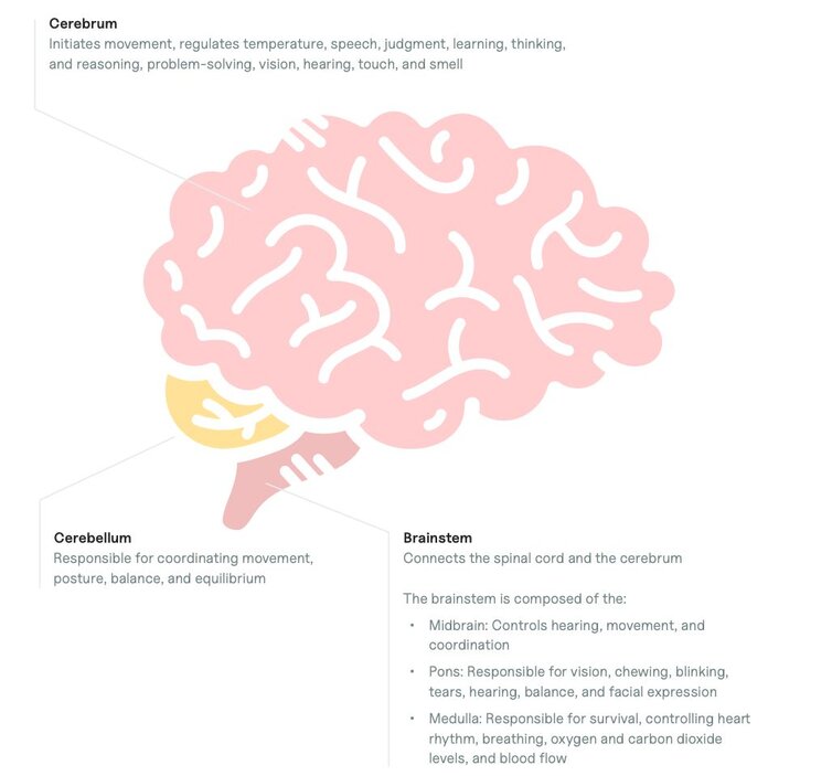
Looking at the brain from a bird's eye perspective, the brain can be split into three sections:
Cerebellum
Located at the back of the head, the cerebellum is responsible for coordinating movement, posture, balance, and equilibrium.
Brainstem
The brainstem is the middle part of the brain, connecting the spinal cord and the cerebrum.
The brainstem is composed of the:
- Midbrain: Controls hearing, movement, and coordination
- Pons: Responsible for vision, chewing, blinking, tears, hearing, balance, and facial expressions
- Medulla: Responsible for survival, controlling heart rhythm, breathing, oxygen and carbon dioxide levels, and blood flow
Cerebrum
The cerebrum is the largest section of the brain, composed of gray matter and white matter in the center.
The cerebral cortex, which is the outer gray matter, is composed of the right and left hemispheres, each controlling opposite sides of the body. These two hemispheres communicate with each other through white matter and nerve pathways, called the corpus callosum.
The cerebrum is responsible for initiating movement, regulating temperature, speech, judgment, learning, thinking, reasoning, problem-solving, vision, hearing, touch, and smell.

Lobes of the brain
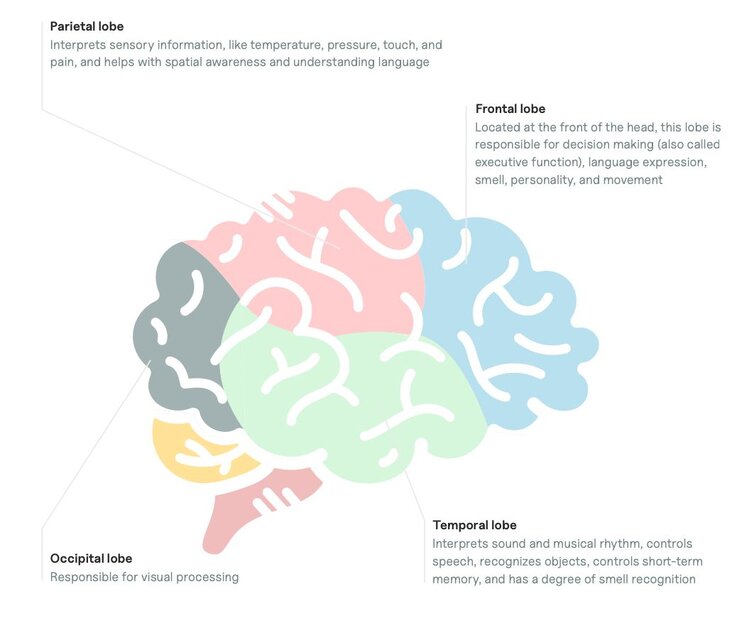
The cerebral cortex can be further broken down into four lobes, each having specific functions:
Frontal lobe
Located at the front of the brain, the frontal lobe is responsible for decision making (also called executive functioning), language expression, smell, personality, and movement.
Parietal lobe
The brain’s parietal lobe interprets sensory information, like temperature, pressure, touch, and pain. It also helps with spatial awareness and language comprehension.
Occipital lobe
The occipital lobe is located at the back of the brain, and it is responsible for visual processing, including color and motion, and memory storage.
Temporal lobe
The temporal lobe interprets sound and musical rhythm, controls speech, recognizes objects, controls short-term memory, and has a degree of smell recognition.

Deeper structures in the brain
The deeper areas of the brain are responsible for many of the responses caused by trauma and mental health conditions. These structures are also responsible for essential bodily functions that can affect our everyday lives.
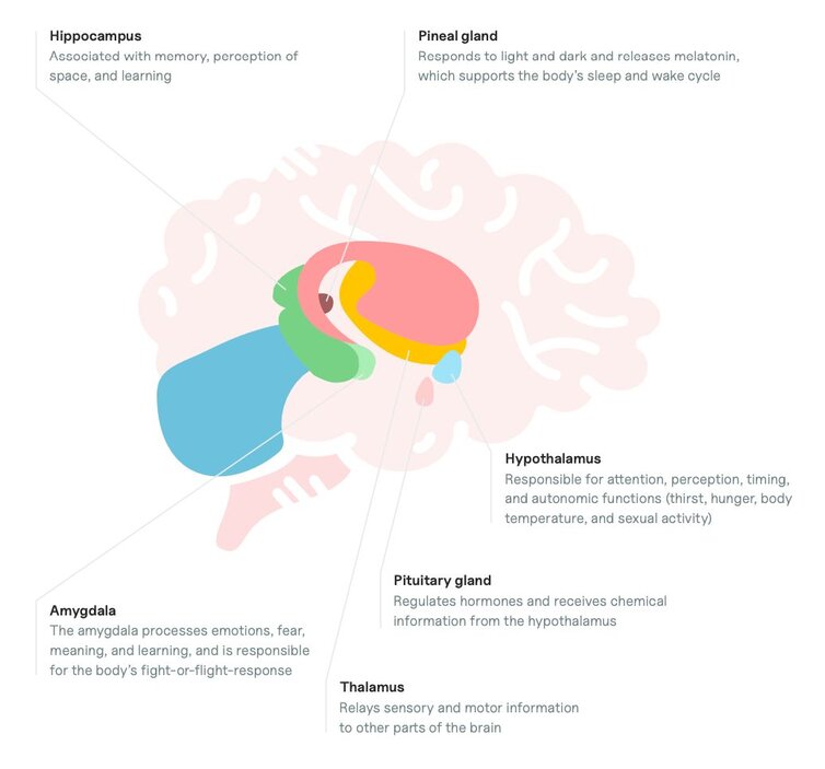
The deeper structures of the brain include:
Amygdala
The amygdala processes emotions, fear, meaning, and learning, and is responsible for the body’s fight-or-flight-response.
Pituitary gland
The pituitary gland regulates hormones and receives chemical information from the hypothalamus.
Thalamus
The thalamus processes all incoming motor and sensory information from a person’s body to their brain, and plays a role in consciousness and alertness.
Hypothalamus
The hypothalamus links a person's nervous system and endocrine system, and is responsible for attention, perception, timing, and autonomic functions (thirst, hunger, body temperature, and sexual activity).
Hippocampus
The hippocampus, located within the temporal lobe, is associated with memory, perception of space, and learning.
Pineal gland
The pineal gland responds to light and dark and releases melatonin, which supports the body’s sleep and wake cycle.
The free brain diagram, labeled with these parts of the brain, can be used as a visual aid for clients to see where each part of the brain is located.

How to use the human brain diagram to educate clients
Using this diagram of a brain with labels can demonstrate the complexity involved in why a person behaves the way they do.
For example, clients may minimize their behaviors and label themselves as “stupid.” They may also struggle to understand why they can’t control a habit, like drinking excessively, despite the negative consequences.
As a therapist, you can use a brain model, labeled with the parts of the brain responsible for these habits, to illustrate the physiological processes that dictate their behavior or emotional state.
Here are some specific examples of concepts or scenarios where you can use a brain function chart:
Clients with post-traumatic stress disorder (PTSD)
Presentation
A client presents with nightmares, sleep disturbances, flashbacks, problems with memory, and feels like they are reliving a traumatic event.
Psychoeducation
Trauma causes a stress response, which can affect the prefrontal cortex, amygdala, hippocampus, and pituitary gland. This triggers the release of stress hormones which fuel the body’s ability to survive, increasing alert and hypervigilant type behaviors.
Explanation of maladaptive behaviors
This stress response can:
- Reduce function in the prefrontal cortex, making it difficult to make decisions, like calling for help or recognizing future stress triggers.
- Cause lasting effects and malfunctioning to the hippocampus and amygdala, making it difficult to recognize threats and mediate an appropriate response. It can also increase startle responses and trigger the release of stress hormones into the body, which may explain why it is often difficult for a person with PTSD to sleep. The production of stress hormones can make them feel on edge and experience difficulty relaxing.
Clients with attention-deficit hyperactivity disorder (ADHD)
Presentation
A client presents with difficulty concentrating, often loses important items, struggles to follow directions and retain information, and gets easily frustrated or angry.
Psychoeducation
Research suggests that there may be atypical interactions between the frontal lobe and information processing centers deeper within the brain in children with ADHD.
Explanation of maladaptive behaviors
The frontal lobe is responsible for judgment, planning, problem solving, attention, time perception, impulse control, motivation, and social interaction, which are all correlated with ADHD symptoms.
Research also suggests that people with ADHD have different levels of neurotransmitters in their brains, such as norepinephrine and dopamine, which are responsible for reward, attention, shifting between tasks, emotions, behavior, movement, and focus.
Using the human brain diagram in your office
You can use a human brain diagram in several ways in your therapy office, such as:
- Frame a brain chart and use it as decor in your private practice
- Stick an image to your whiteboard to use in-session (or use a virtual whiteboard for telehealth sessions)
- Save the brain diagram labeled to your computer or EHR to use in virtual sessions
- Have printed versions available for clients to write on and take home
How SimplePractice streamlines running your practice
SimplePractice is HIPAA-compliant practice management software with everything you need to run your practice built into the platform—from booking and scheduling to insurance and client billing.
If you’ve been considering switching to an EHR system, SimplePractice empowers you to streamline appointment bookings, reminders, and rescheduling and simplify the billing and coding process—so you get more time for the things that matter most to you.
Try SimplePractice free for 30 days. No credit card required.

